Study of Meiosis Using Permanent Slides
Study of Meiosis Using Permanent Slides
Objective: To observe and identify the stages of meiosis using prepared permanent slides, and to understand the process of meiosis and its role in sexual reproduction.
Introduction:
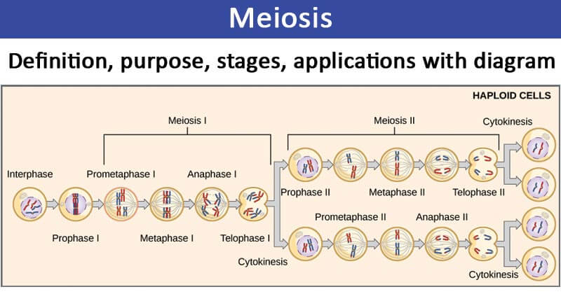
Meiosis is a type of cell division that reduces the chromosome number by half, resulting in four genetically diverse haploid cells from one diploid parent cell. It is crucial for sexual reproduction and occurs in two main stages: meiosis I and meiosis II. Permanent slides of various organisms are often used to study meiosis, as they provide a clear view of the different stages.
Materials and Reagents:
1. Permanent Slides of Meiosis Stages: Prepared slides with stained specimens showing various stages of meiosis (e.g., from plant or animal tissues).
2. Microscope: Equipped with objective lenses (4x, 10x, 40x, and 100x) and an eyepiece.
Procedure:
1. Preparing the Microscopy Setup:
1. Select a Slide: Choose a permanent slide that has been prepared to show the stages of meiosis. Common slides might include those prepared from plant tissues (e.g., onion flowers) or animal tissues (e.g., testis of mammals).
2. Setting Up the Microscope: Place the permanent slide on the microscope stage. Ensure that the light source is adjusted for optimal illumination.
2. Observing the Stages of Meiosis:
1. Initial Examination: Start with the lowest magnification objective lens (e.g., 4x or 10x) to locate the area of interest on the slide.
2. Fine Focusing: Switch to a higher magnification objective lens (e.g., 40x or 100x) for a detailed view of the stages of meiosis.
3. Identifying the Stages:
⮚ Prophase I: Look for homologous chromosomes pairing up (synapsis) and forming tetrads. Crossing-over (exchange of genetic material between homologous chromosomes) may also be visible.
⮚ Metaphase I: Observe the tetrads aligned along the metaphase plate.
⮚ Anaphase I: Identify the separation of homologous chromosomes, with each chromosome moving toward opposite poles.
⮚ Telophase I: Look for the formation of two new nuclei around each set of chromosomes, resulting in two haploid cells.
⮚ Prophase II: In the second meiotic division, look for the formation of a new spindle apparatus in each haploid cell.
⮚ Metaphase II: Observe chromosomes aligned along the metaphase plate in each haploid cell.
⮚ Anaphase II: Identify the separation of sister chromatids toward opposite poles
⮚ Telophase II: Look for the formation of four genetically diverse haploid cells.
4. Documenting Observations:
o Draw or take images of each stage of meiosis observed. Label the stages and any notable features.
Results:
Different stages of meiosis was observed and studied about the structure and alignment of chromosomes.
Conclusion:
Studying meiosis using permanent slides provides a clear view of the complex process of cell division that is essential for sexual reproduction. By observing and identifying the stages of meiosis, one can gain a deeper understanding of how genetic material is distributed and how genetic diversity is generated.

Comments
Post a Comment