Squash Preparation of Polytene Chromosomes
Title: Squash Preparation of Polytene Chromosomes
Objective:
To prepare and observe polytene chromosomes from the salivary glands of Drosophila melanogaster (fruit fly) or Chironomus larvae, and to study their unique banding patterns and structure.
Theory:
Polytene chromosomes are giant chromosomes found in the salivary glands of dipteran insects like Drosophila melanogaster. They result from repeated rounds of DNA replication without cell division, leading to the formation of large, multi-stranded chromosomes. These chromosomes exhibit distinct banding patterns due to variations in chromatin condensation, which can be observed under a microscope. Studying polytene chromosomes provides insights into chromosome structure, gene activity, and chromosomal aberrations.
Materials Required:
∙ Drosophila melanogaster larvae or Chironomus larvae
∙ Forceps
∙ Needles
∙ Microscope slides
∙ Cover slips
∙ Aceto-orcein stain/ Aceto-carmine stain
∙ Distilled water
∙ Dropper or pipette
∙ Light microscope
Procedure:
1. Obtain third-instar larvae of Drosophila melanogaster or Chironomus from a culture and place a larva on a clean microscope slide
2. Head portion of the larvae was dissected using a needle and remaining portion was discarded.
3. It was squashed well with a needle.
4. Carefully add a few drops of stain to the salivary glands on the slide and incubate for 5-10 minutes. This will stain the chromosomes and enhance the visibility of the banding pattern.
5. Gently place a cover slip over the stained salivary glands.
6. Use the blunt end of a pencil or another smooth object to gently press down on the cover slip, squashing the glands to spread out the chromosomes.
7. excess stain was blot dried using a blotting paper
8. Observe the slide under a light microscope at high magnification (400x or higher).
9. Focus on the polytene chromosomes, which should appear as large, banded structures. The bands represent regions of tightly packed DNA, while the interbands are less condensed.
Observations:
∙ Polytene Chromosomes: Look for the distinct, banded pattern of polytene chromosomes. The chromosomes may show puffing, which is an indication of gene activity.
Results:
∙ The preparation should reveal polytene chromosomes with clear, alternating dark and light bands. Any puffs or swelling on the chromosomes indicate active transcriptional regions.
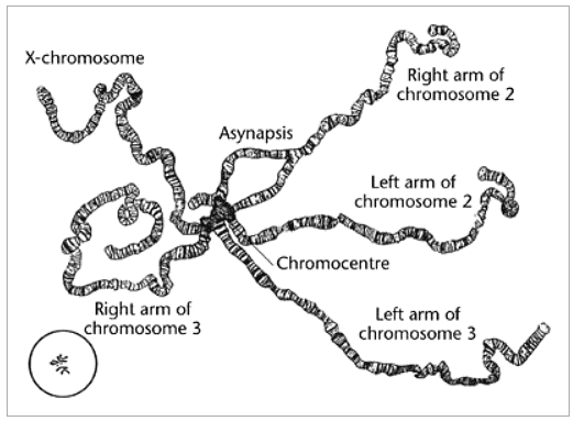

Conclusion: This experiment allows for the observation of polytene chromosomes, providing insight into their structure and function. The banding pattern observed is crucial for understanding chromosomal organization and gene activity.
Precautions:
∙ Handle the larvae carefully during dissection to avoid damaging the salivary glands. ∙ Ensure the squash is done gently to avoid breaking the chromosomes. ∙ Use fresh stains and reagents for clear visibility of the chromosome bands.


Comments
Post a Comment