Observation of Barr body in Human Buccal Epithelial Cells
Title: Observation of Barr body in Human Buccal Epithelial Cells
Objective:
To observe and identify the Barr body in buccal epithelial cells and understand its significance in sex determination and X-chromosome inactivation.
Theory:
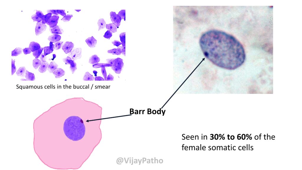
The Barr body is an inactivated X chromosome found in the nuclei of female cells. It appears as a densely stained structure at the periphery of the nucleus. This phenomenon is a result of X chromosome inactivation, where one of the two X chromosomes in females becomes transcriptionally inactive to balance the dosage of X-linked genes between males and females. The Barr body can be used as a cytological marker for determining the sex of an individual. This darkly stained structure is named after Dr Murray Barr who was a Canadian physician who along with his student Dr Bertram way back in 1948 identified this structure.
Materials Required:
∙ Sterile buccal swab/tooth pick
∙ Microscope slides
∙ Cover slips
∙ Acetocarmine stain/Giemsa/Toluidine blue
∙ Distilled water
∙ Dropper or pipette
∙ Light microscope
Procedure:
1. Use a sterile buccal swab/tooth pick to gently scrape the inner lining of the cheek to collect epithelial cells after washing mouth thoroughly with water.
2. Smear the collected cells onto the centre of a clean microscope slide and allow the slide to air dry completely.
4. Add a few drops of acetocarmine stain and allow it to stain for 5-10 minutes.
7. Gently rinse off the excess stain with distilled water by keeping in a slanted position.
8. Eventually, water can be blotted using tissue paper.
9. Place a cover slip over the stained sample.
10. Observe the slide under a light microscope at high magnification.
11. Look for the Barr body, which appears as a small, darkly stained mass near the nuclear membrane.
Observations:
∙ Barr Body Presence: Identify and note the presence of a small, round or oval-shaped, darkly stained structure at the edge of the nucleus in some cells.
∙ Barr Body Absence: In some cells, particularly in male samples, there will be no Barr body present.
Results:
∙ The presence of a Barr body indicates a cell from a female, as it confirms the presence of two X chromosomes with one being inactivated.
∙ The absence of a Barr body indicates a male cell, which typically has only one X chromosome.
Conclusion: This experiment demonstrates the concept of X-chromosome inactivation and allows for the identification of the Barr body as a marker of sex chromatin. The Barr body serves as an important tool in cytogenetics for sex determination.
Precautions:
∙ Ensure the cells are spread evenly on the slide to avoid overlapping, which can obscure the Barr body.
∙ Handle stains and fixatives carefully as they are hazardous.

Comments
Post a Comment