Karyotyping
Karyotyping is a laboratory technique used to visualize and analyze the number, size, and shape of chromosomes in a cell. This process is essential for detecting chromosomal abnormalities that can lead to genetic disorders. Karyotyping is typically performed on cells in the metaphase stage of mitosis when chromosomes are most condensed and easily visible under a microscope.
Steps in Karyotyping
1. Sample Collection:
o Sources: Common sources of cells for karyotyping include blood (white blood cells), bone marrow, amniotic fluid (for prenatal testing), or tissue samples.
o Cell Culture: The collected cells are cultured in a laboratory to increase their number and ensure that a sufficient number of cells are in the metaphase stage.
2. Cell Harvesting:
o Arresting Mitosis: Cells are treated with a mitotic inhibitor like colchicine, which stops cell division at metaphase, where chromosomes are most visible. o Hypotonic Treatment: Cells are then placed in a hypotonic solution, causing them to swell. This spreads the chromosomes within the cell, making them easier to observe.
3. Chromosome Staining:
o Giemsa Stain (G-banding): The most common staining technique is G-banding, which uses Giemsa stain to produce a distinct banding pattern on chromosomes. These bands represent areas of different densities of chromatin, allowing for the identification of each chromosome.
4. Microscopic Examination:
o Slide Preparation: The stained cells are placed on a microscope slide, and the chromosomes are observed under a light microscope.
o Photomicrography: High-resolution images of the chromosomes are taken. 5. Karyotype Analysis:
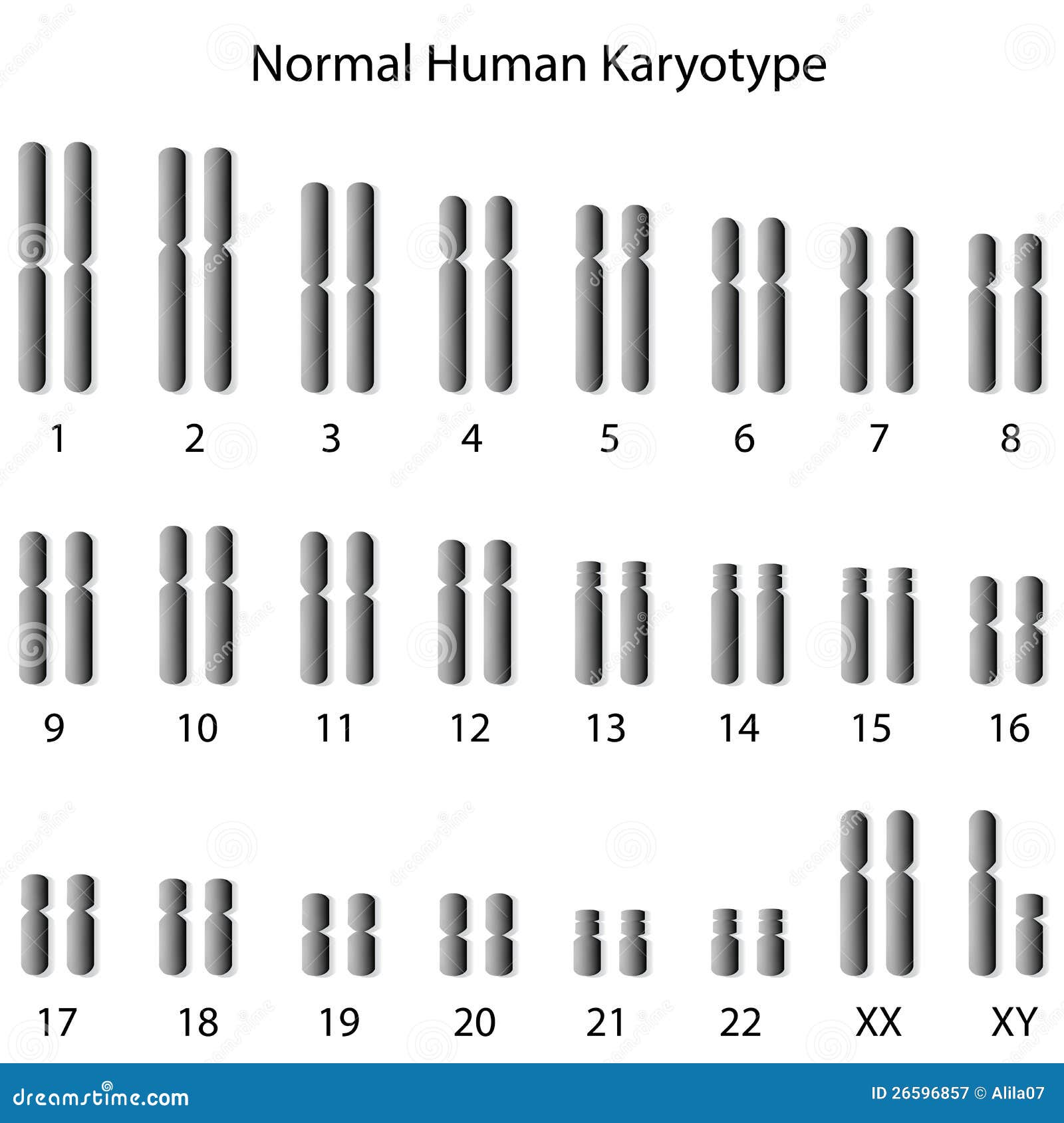
o Arrangement: Chromosomes are arranged in pairs according to their size, banding pattern, and centromere position, from largest to smallest. The sex chromosomes (X and Y) are usually placed at the end.
o Karyogram: The resulting image, known as a karyogram, shows the complete set of chromosomes in an individual’s cells. A typical human karyotype consists of 46 chromosomes, arranged into 23 pairs.

Applications of Karyotyping
1. Diagnosis of Genetic Disorders:
o Aneuploidy: Karyotyping can detect abnormalities in chromosome number, such as trisomy (an extra chromosome) or monosomy (a missing chromosome). For example:
▪ Down Syndrome: Trisomy 21, where there is an extra copy of chromosome 21.
▪ Turner Syndrome: Monosomy X, where there is only one X chromosome.
o Structural Abnormalities: Karyotyping can also identify structural changes in chromosomes, such as:
▪ Translocations: When a segment of one chromosome is transferred to another chromosome.
▪ Deletions: Loss of a chromosome segment.
▪ Duplications: Extra copies of a chromosome segment.
▪ Inversions: A segment of a chromosome is reversed end to end.
2. Prenatal Testing:
o Amniocentesis: Karyotyping of fetal cells obtained from amniotic fluid can detect chromosomal abnormalities before birth.
o Chorionic Villus Sampling (CVS): Another prenatal test where cells from the placenta are analyzed.
3. Cancer Research and Diagnosis:
o Chromosomal Aberrations: Certain cancers are associated with specific chromosomal changes. For example, the Philadelphia chromosome is a
translocation between chromosomes 9 and 22, found in chronic myeloid leukemia (CML).
4. Infertility Investigations:
o Chromosome Analysis: Karyotyping can help identify chromosomal abnormalities that might be causing infertility or recurrent miscarriages.
5. Species Identification and Evolutionary Studies:
o Comparative Karyotyping: Karyotypes can be compared between species to study evolutionary relationships.
Limitations of Karyotyping
∙ Resolution: Karyotyping cannot detect very small chromosomal changes, such as microdeletions or duplications. More advanced techniques like fluorescence in situ hybridization (FISH) or comparative genomic hybridization (CGH) are required for higher-resolution analysis.
∙ Time-Consuming: The process of culturing cells and preparing karyotypes can be time consuming, often taking several days.
Conclusion
Karyotyping remains a fundamental tool in genetics, providing valuable insights into chromosomal structure and abnormalities. It plays a crucial role in diagnosing genetic disorders, understanding cancer, and conducting prenatal screening. Despite its limitations, karyotyping continues to be widely used due to its ability to visualize the entire chromosome set in a single analysis.

Comments
Post a Comment